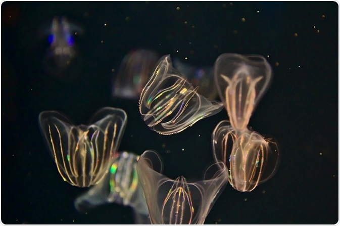
The occurrence of bioluminescence is extremely diverse in nature. However, it is found to be rare if we calculate their presence across the entirety of the species. Although fluorescence and bioluminescence occur in aquatic and terrestrial organisms, they are widespread among aquatic organisms to attract mates, to find prey, or to protect themselves from their enemies.

© Karen Adamczewski/Shutterstock.com
Earlier studies have shown evidence that glowing light was produced in certain biological organisms such as jellyfish and fireflies. A similar light emitting method is seen in deep sea fish to attract and consume the prey. About 90% of deep ocean animals are considered to be luminescent. When compared with the ocean, the occurrence of bioluminescence in freshwater and land is not common.
Bioluminescence
Around 50% of jellyfish are estimated to be bioluminescent. Deep in the sea, luminescent jelly fish are seen in greatest diversity – Sea pens (Phylum: Cnidaria; Class: Anthozoa), Medusae (Phylum: Cnidaria; Subphylum: Medusozoa), Siphonophores (Phylum: Cnidaria; Class: Hydrozoa) generate several particles, resembling a glowing chain for puzzling the predator; some other types of jelly fish produces a slime that glows to make its predator stick to them or produce glowing decoys while releasing the tentacles.
Genes of green fluorescent proteins (GFP) were identified in nearly 100 animal species. Some species have more than six gene copies and generate a broad range of colors. Yet all these species fall into one of the three phyla: Cnidaria or Chordata or Arthropoda.
Phylum Cnidaria
In 1962, GFP was first identified in the jellyfish Aequorea victoria. Researcher Osamu Shimomura discovered GFP under ultraviolet radiation. The Royal Swedish Academy of Sciences awarded the Nobel Prize (Chemistry) to Osamu Shimomura, Martin Chalfie, and Roger Y. Tsien in 2008 for discovering and developing GFP.
In Aequorea victoria jellyfish, the protein aequorin is present in the photo organs. When the protein binds with calcium, a blue light is produced. GFP absorbs the blue light completely and turns it into green light. The presence of 238 amino acids in Aequorea GFP constitutes its polypeptide size of about 27kDa. The GFP molecules have maximum excitation in the ultraviolet region; 395 nm is the major excitation peak; 475 nm is the minor excitation peak; and 508 nm is the emission peak.
Subsequently, GFP was discovered in the genus Obelia. In 1970, researchers were able to track the genes creating fluorescent proteins in Obelia – a type of jelly fish. Other than green, cyan and yellow fluorescent protein colors were found in Obelia medusa jellyfish.
Among the phylum cnidaria, GFP-like fluorescent proteins have been discovered in corallimorpharians, hydroids, corals, pennatulids, and anemones.
Class Anthozoa possesses most of the species expresses fluorescent proteins.
Reef-building coral (Scleractinia), the subgroup of the Class Anthozoa, has green fluorescent proteins and it is localized in unique anatomical patterns. In a study involving coral specimens collected across 33 species from the family, Poritedae, Siderasteidae, Acroporidae, Faviidae, Fungiidae, Merulinidae, Mussidae, Oculinidae, and biofluorescence was present in 28 species.
GFP was found to be dominant among the fluorescent emissions contributing to 480–520 nm. The fluorescent proteins were found to be concentrated on the tentacles in most cases and fluorescence was not exhibited in other coral areas. The intensity of the fluorescence varied across the 8 families yet the fluorescent patterns remained constant.
It was found that the GFP from the species Renilla kollikeri and Renilla reniformis, sea pansy, is advantageous when compared with Aequorea GFP. The emission spectrum resembles the same in both the species; however, the absorption spectrum of GFP in the genus Renilla is very diverse. For exciting Renilla GFP, higher wavelength is utilized.
The excitation peaks of the GFP molecules in Renilla are at 498 nm and 470 nm; absorption is in the range of 320–390 nm. This very little region of absorption is used in applications such as fluorescence microscopy. The extinction coefficient is higher in Renilla GFP when compared with Aequorea GFP making it possible to use in vivo expression. Renilla GFP shows higher stability upon subjecting to pH extremes paving way to several in vitro applications.
Phylum Arthropoda
Studies have found many fluorescent proteins in the sub class, Copepoda (family: Pontellidae). The overexpression of GFP, ppluGFP2, from the species Pontellina plumata in mammalian cells has led to microcrystal formation which ultimately ruptures the membranes. So, their applications are restricted.
Phylum Chordata
Another study has found GFP in 3 species of the Phylum Chordata (genus: Branchiostoma) – B. floridae, B. lanceolatum, and B. belcheri. The AmphiGFP (green fluorescence) from B. floridae is concentrated on the anterior end and exhibits strong relationship with copepod GFPs than cnidarians GFPs.
Phylum Ctenophora
In comb jellies belonging to the phylum Ctenophora, GFP-type fluorescent proteins were discovered. The bright flashes of the comb jellies – the ctenophore proteins exhibit reversible photoactivation property. In this rare property, upon exposing to light, the fluorescent emission will become bright and gradually reduced to a non-fluorescent state.
 Study shows cutaneous touch neuronal cells to be critical for sexual receptiveness