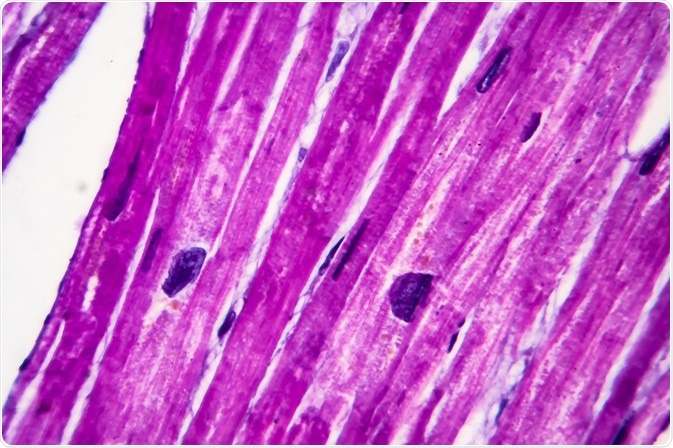
The mammalian heart is composed of different cell types with different functions, which are not all understood in detail. In order to characterize the physiology and type of cell lines in laboratory tests, cell extraction methods are required.
 Image Credit: Kateryna Kon / Shutterstock.com
In a recent method publication, scientists from Switzerland and Poland have described a cell sorting procedure for collagen-producing heart fibroblasts from the hearts of laboratory mice by using fluorescence-activated cell sorting (FACS) with cell type-specific, fluorescently labeled antibodies.
This research has been published in the journal Frontiers in Cardiovascular Medicine.
Investigating the cellular composition of the heart is important for understanding cardiac health and disease. Meanwhile, not all cell types have yet been identified. In order to characterize the cells in detail, heart tissue is commonly disintegrated by enzymatically removing the extracellular matrix, and identifying cell types of interest-based on different cell surface markers.
For collagen-producing fibroblasts, no such markers had been known. Instead, transgenic mouse lines had been used in which these particular fibroblasts expressed the fluorescent protein EGFP. Since the fibroblasts had been inherently fluorescent, cell sorting could easily differentiate them from other cardiac cells, and fibroblasts could be examined in in vitro cell cultures.
However, in transgenic cell lines, there is always the potential that the non-native expression of a protein like EGFP changes the cellular physiology, and non-transgenic cell lines would thus be preferable.
Researchers from Switzerland and Poland, led by Assoc. Professor Gabriela Kania, have now shown that the collagen-producing fibroblasts in mouse hearts can alternatively be identified in fluorescence -activated cell sorting (FACS) based on tagging two cell surface markers with fluorescent antibodies.
Identifying the right surface markers
The scientists started their method development from transgenic mice with an EGFP fluorescent label. The gene for the EGFP protein was controlled by an expression-promoter that naturally controls a collagen-production gene, thus making the EGFP expression representative for collagen-producing fibroblasts.
Having identified the collagen-producing fibroblasts, the researchers tested the binding of different antibodies that are specific for particular surface proteins and identified that the EGFP-positive cell population was also positive for the cell-surface marker gp38. In addition, they found that the EGFP-positive, gp38-positive fibroblasts were negative for three other surface markers called Ter119, CD45, and CD31.
As a result, the scientists had identified that collagen-producing cardiac fibroblasts form a unique population in flow cytometry with high fluorescence from the gp38 tagging, with separation from other cell lines based on their lack of fluorescence from antibodies that are specific for Ter119, CD45, and CD31.
Physiological testing of fibroblasts
The investigators next demonstrated that fibroblasts isolated by their FACS method could be taken into in vitro cell culture for physiological assays. Here, they did not use a transgenic EGFP mouse line, but instead a wild-type line isolated with their new FACS method.
By testing for cell line-specific cell surface markers, they demonstrated that after extended in vitro culture of the isolated cell line (4 passages), the cells were still similar to their ancestor cells, but also showed changed patterns of some surface markers.
As the study authors note, their FACS method for the isolation of fibroblasts can be useful for the analysis of hearts in experimental models of cardiac diseases. As the method is efficient for fibroblast isolation, and fairly easy to carry out, it can be widely used in standard laboratories and overcomes the need for transgenic mouse lines where fibroblasts are inherently fluorescent.
The focus on collagen-producing fibroblasts is thereby of particular interest, as unbalanced formation of collagen in the heart is the camajor use of heart disease, fibrosis and improved understanding of the collagen-forming cell lines could help to identify an appropriate therapeutic approach against it.
Sources
Stellato M et al. Identification and Isolation of Cardiac Fibroblasts From the Adult Mouse Heart Using Two-Color Flow Cytometry. Front Cardiovasc Med 2019, 6, 105; DOI: 10.3389/fcvm.2019.00105.
The authors note that animal experiments were approved by the cantonal veterinary office of Zurich (Switzerland).
Further Reading
 Clinically relevant biomarker strategies in drug development