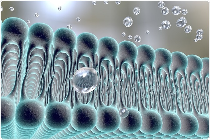
Ferroptosis is a form of programmed cell death, distinct from apoptosis, that was identified in 2012. The process is driven by the iron-dependent oxidative degeneration of lipids.
 Image Credit: Kateryna Kon / Shutterstock
Deregulated ferroptosis is thought to cause neurodegenerative disease and ischemic injury. There is also evidence that ferroptosis plays a fundamental role in the regulation of growth and proliferation of certain cancer cells. By learning more about ferroptosis, scientists hope to develop new therapeutic anticancer strategies.
Mechanisms of ferroptosis
Ferroptosis causes cell death through the accumulation of iron-dependent lipid reactive oxygen species. Free radicals cause degradation of lipid molecules by the removal of electrons through oxidation.
The process is dependent on intracellular iron as the accumulation of iron acts as a catalyst for converting peroxides into free radicals. The oxidative degradation of lipids occurs when there is depletion of the antioxidant glutathione and a loss of activity of the lipid repair enzyme glutathione peroxidase 4 (GPX4). The lipid peroxidation then leads to cell membrane denaturation.
The mechanism underpinning cell death once ferroptosis is triggered remains undefined. Accumulation of redox-active iron, glutathione depletion and lipid oxidation are presumed to occur simultaneously as the prevention of one critical event halts ferroptosis.
A positive feedback loop may amplify these events with the combined accumulation of iron toxicity, antioxidant depletion, and membrane damage producing cell death. The sensitivity of a cell towards ferroptosis is correlated with lipid metabolism.
Polyunsaturated fatty acids are susceptible to lipid peroxidation with their abundance and localization determining the amount of lipid peroxidation that occurs within the cell and therefore the extent of ferroptosis.
Why does ferroptosis exist?
The role of ferroptosis in normal physiological function is currently unknown but there is emerging evidence that the process may have a tumor suppressor function by removing cells compromised by environmental stress or lacking access to required nutrients.
Studies have shown that small molecules can induce ferroptosis, inhibiting tumor growth and enhancing the sensitivity of chemotherapeutic drugs. Induced ferroptosis may therefore have a future role in cancer therapy.
Nonetheless, there are still questions to be answered about how ferroptosis is genetically programmed, along with defining roles during development and tumorigenesis. Moreover, whether ferroptosis is adaptive or a result of a biochemical failure in cells is still unknown.
One theory suggests that though the incorporation of polyunsaturated fatty acids into cell membranes during evolution allowed for more complex physiological functions, it produced a susceptibility to lipid peroxidation.
Ferroptosis and pathology
There appears to be a connection between ferroptosis and degenerative diseases because high iron loads, lipid damage, and glutathione consumption are features of Parkinson’s disease, Huntington’s disease, and Alzheimer’s disease.
Ferroptosis can cause inflammation in the brain through the degradation of neurons and the resulting release of lipid metabolites. Experiments on mice models found decreased GPX4 levels produced cognitive impairment and neurodegeneration of hippocampal neurons.
Brain iron levels are known to increase with age and in degenerative diseases, suggesting a potential increased risk of ferroptosis with age.
Cell death through ferroptosis is a primary contributor to liver, kidney, brain, and heart injuries tested through mouse models. During muscle injury the breakdown of muscle products enters the circulation and can lead to acute organ damage such as kidney failure.
The iron released from the heme of myoglobin promotes ferroptosis and therefore damages kidney cells. Ferroptosis inhibitors, such as ferrostatins and liproxstatins, have been proven to protect organ function from ischemic injury. Studies have also found that ferroptosis inhibitors provide protection in models of degenerative brain disorders and hemorrhagic brain injury.
Sources:
- Dixson et al. 2012. Ferroptosis: An Iron-Dependent Form of Non-Apoptotic Cell Death, Cell, 149, pp. 1060-1072.
- Cao, JY. & Dixon, S.J. 2016. Mechanisms of ferroptosis, Cellular and Molecular Life Sciences, 73, pp. 2195-2209.
- Bertrand, R.L. 2017. Iron accumulation, glutathione depletion, and lipid peroxidation must occur simultaneously during ferroptosis and are mutually amplifying events, Medical Hypotheses, 101, pp. 69-74.
- Yang, W.S. & Stockwell, B.R. 2016. Ferroptosis: Death by Lipid Peroxidation, Trends in Cell Biology, 26, pp. 165-176.
- Stockwell, B.R. et al. 2017. Ferroptosis: A Regulated Cell Death Nexus Linking Metabolism, Redox Biology, and Disease, Cell, 171, pp. 273-285.
Further Reading
 Researchers explore the possibility of treating lung cancer by targeting telomeres